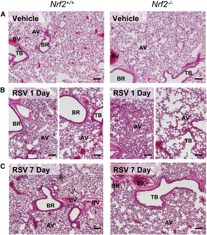
Figure 3.
Nrf2 deficiency exacerbated lung histopathologic phenotypes of respiratory syncytial virus (RSV) infection. Representative light photomicrographs of lung sections from Nrf2+/+ and Nrf2−/− mice (n = 3–5/group) of vehicle controls (A) or 1 day (B) and 7 days (C) after infection stained with H&E. Increased airway cellularity in proximal sections (left panels) and loss of alveolar structure in distal sections (right panels) was marked in Nrf2−/− mice relative to Nrf2+/+ mice at 1 day. Bronchial and alveolar epithelial proliferation (hyperplasia) and alveolar vacuolization remained more obvious in Nrf2−/− mice than in Nrf2+/+ mice by 7 days. AV = alveoli; BR = bronchi or bronchiole; BV = blood vessel; TB terminal bronchiole. Bars indicate 100 μm.

