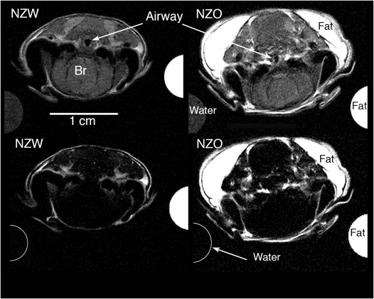
Figure 1.
Four axial magnetic resonance images were obtained from the Dixon protocol at the level of the hypopharynx (caudal pharynx) in representative New Zealand White (NZW) and New Zealand Obese (NZO) mice. The top row shows proton-weighted images (for soft tissue contrast and segmentation) and the bottom row shows fat-weighted images (for fat segmentation and threshold analysis). The half circles on left and right of each image show the partial cross-section of the water and mineral oil (fat) phantoms. In the top row both the water and fat cross-sections can be seen at the resonant frequency for water. In the bottom row, only the fat oil phantom has a white intensity at the resonant frequency for fat; the water phantom shows the water signal intensity is completely suppressed. The use of water and oil phantoms provided calibration signals for the threshold analysis. The airway caliber in the top row (proton weighted images) is larger in the NZW mice compared with NZO mice, and there is more fat (in the fat-weighted images, bottom row) in the NZO mice compared with NZW mice. The upper airway is not well delineated in the fat-weighted images (bottom row) because the soft tissues, immediately adjacent to the airway, have little fat content.

