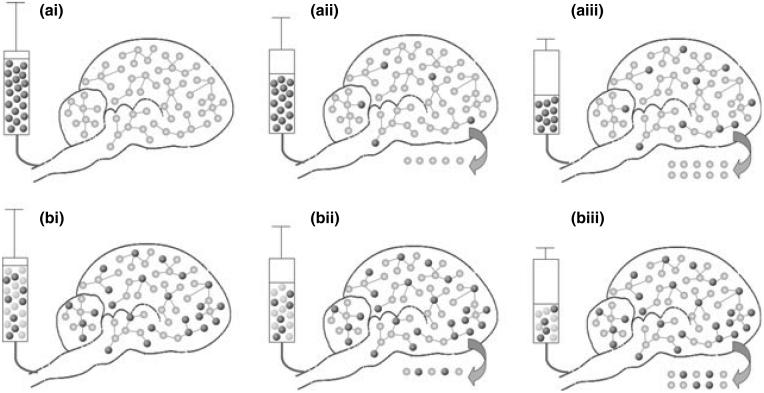
Fig. 1.
Schematic illustration of the effect of label turnover on glycogen quantification. (a) Brain glycogen is labeled by infusing a highly enriched solution of 13C-labeled Glc (dark spheres) into an unlabeled rat (a-i) femoral vein. Light spheres represent 12C-Glc (so-called unlabeled Glc). An increase in 13C signal can be observed over time (represented by an increase in the number of dark spheres), even in the absence of changes in brain glycogen concentration (the total number of spheres does not change over time). The 13C signal augmentation is thus because of an increase in glycogen IE over time. (b) The new method uses long term administration of 13C-labeled Glc prior to the measurement to achieve significant brain glycogen IE. After measuring this IE in vivo (using NAA IE as an indirect method), the same IE of the infused Glc solution is matched to that of brain glycogen IE and the 13C-NMR glycogen measurement is started. By subsequently infusing a Glc solution whose IE is similar to that of glycogen, glycogen IE remains stable over time and therefore no change in signal intensity is observed unless the absolute glycogen concentration changes (total number of spheres). Due to glycogen turnover, represented by the arrow, the glycogen concentration in the rat brain remains constant (schematized here as a constant number of spheres) despite the infusion of glucose. N.B.: 13C natural abundance (1.1%) has been neglected (especially in a-i).

