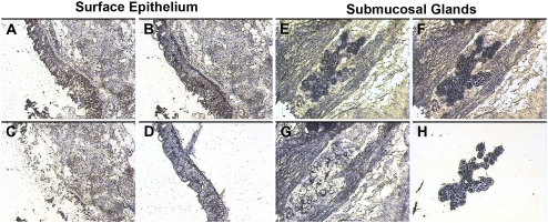
Figure 1.
Laser capture microdissection (LCM) of surface epithelia and submucosal glands (SMG). Representative images from the tissue isolation process are depicted for surface epithelia (left, A–D) and submucosal glands (right, E–H). A and E represent respective tissues before dissection. Captured cells attached to the LCM cap film are demonstrated for surface epithelia (B) and submucosal glands (F). After removal of the LCM cap, unselected tissue remains behind (C and G). The LCM process yields populations of surface epithelial cells (D) and submucosal gland cells (H) on the LCM cap film.

