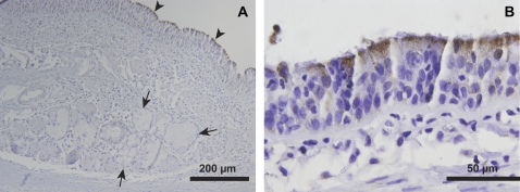
Figure 3.
Mitochondrial staining is greater in conducting airway surface epithelial cells compared with submucosal gland cells. Immunohistochemical staining of bronchus tissue is presented for isocitrate dehydrogenase (IDH3G), a ubiquitous Krebs cycle enzyme with known localization to the mitochondrial matrix. (A) IDH3G immunostaining is highly enriched in surface epithelial cells (arrowheads), with less intense staining observed in SMG epithelial cells (arrows). (B) IDH3G staining was most intense immediately below the apical surface of ciliated epithelial cells. Representative of n = 3 human donor bronchi examined.

