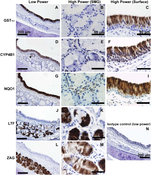
Figure 4.
Immunohistochemical localization of selected gene products confirms the microarray results. (A) GST-α is widely expressed in the surface epithelium. (B) Staining was observed very rarely in serous and mucus submucosal glands. (C) The most intense staining was observed at the apical surface of ciliated epithelial cells. (D) CYP4B1 was specifically observed on a subset of surface epithelial cells, with additional staining in cells lining the gland duct (arrowhead). (E) Under higher power, CYP4B1 was absent in the submucosal glands. (F) Surface CYP4B1 immunostaining was most intense near cilia, which could be consistent with the prediction that CYP4B1 is secreted. Minimal immunostaining was observed in the basal layer of surface epithelial cells. (G) Expression of CYP4B1 declined in the more distal portions of submucosal gland ducts (data not shown). NQO1 staining was widely expressed in the surface epithelium, consistent with previous studies (25). (H) NQO1 was not detectable in submucosal gland epithelia, but light staining was observed in interstitial cells. (I) The basal layer of surface airway epithelial cells was deficient in NQO1 expression. Surface epithelial staining within columnar cells appeared cytoplasmic, with a bias toward apical expression. NQO1 immunostaining was also present in vascular endothelial cells and in chondrocytes of bronchial cartilage (data not shown). (J and K) LTF is expressed highly in the serous demilunes, consistent with earlier observations (26). (L and M) ZAG expression was intense in the vast majority of acinar gland cells, and appeared to be present in both serous and mucus cell types. A representative tissue section stained with isotype control antibody is presented in panel N.

