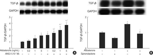
Fig. 2.
Aldosterone stimulates TGF-β1 mRNA expression in mesangial cells. (A) Cells were incubated with various dose of aldosterone (5 ng/mL) or angiotensin II (10-7 M) or a combination of aldosterone and angiotensin II for 16 hr. *p<0.05 vs. untreated control. †p<0.05 vs. angiotensin II-treated cells. (B) Cells were pre-incubated with or without spironolactone (10-9 M) for 60 min and then incubated with aldosterone (5 ng/mL) for 16 hr. *p<0.05 vs. untreated control. †p<0.05 vs. aldosterone-treated cells. In both (A) and (B), Upper panel, the autoradiograph is a representative of five independent experiments with similar results. Lower panel, the optical density of autoradiographic signals was quantified and calculated as the ratio of TGF-β1 to GAPDH mRNA. Results are expressed as fold increase over untreated (represented as 1) in densitometric arbitrary units. Each value represents the mean±SEM.

