Fig. 1.

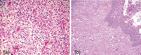
Histological examination of periapical lesions with haematoxylin and eosin stains. (a) Periapical granulomas, which were rich in blood vessels. (b) Radicular cysts with epithelium-lined tissues. Original magnification: ×100 (a) or ×50 (b).

Histological examination of periapical lesions with haematoxylin and eosin stains. (a) Periapical granulomas, which were rich in blood vessels. (b) Radicular cysts with epithelium-lined tissues. Original magnification: ×100 (a) or ×50 (b).