
Fig. 2.
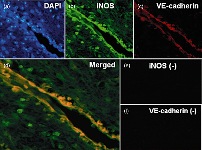
Two-colour immunofluorescence image analysis of periapical granulomas. (a) Nuclear counterstains using 4,6-diamine-2-phenylindole. (b) Inducible nitric oxide synthase (iNOS) monoclonal antibody. (c) Vascular endothelial (VE)-cadherin polyclonal antibody. (d) Merged images. (e) Negative controls for iNOS. (f) Negative controls for VE-cadherin. Original magnification: ×100 (a–c, e, f) and ×200 (d).