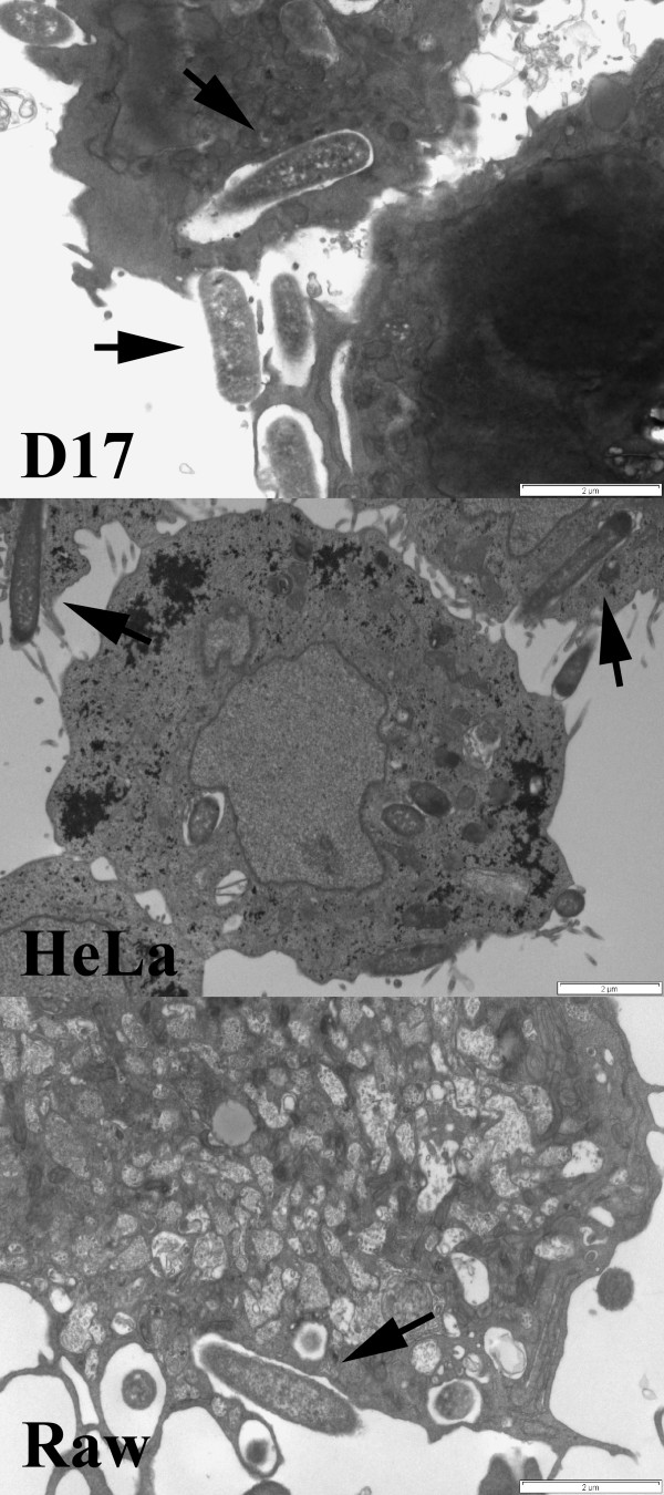
Figure 2.
Transmission Electron Microscopy shows recombinant invasive E. coli similarly engulfed by non-professional phagocytic cells (D17, HeLa) and phagocytic cells (Raw). The osteosarcoma cell line D17, epithelial cell line HeLa, and macrophage cell line Raw were incubated with recombinant invasive E. coli (MOI 10) for 3 hours, washed, fixed and processed for TEM. Image demonstrates each cell line engulfing E. coli (arrows) with lamellipodia. Scale bar indicates 2 microns.

