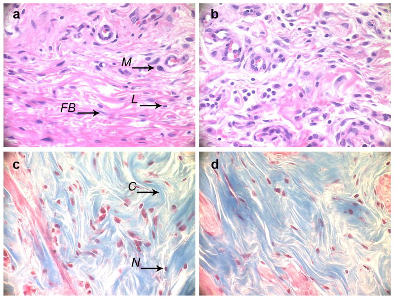
Figure 10.

Histological analysis. H&E (a, b) and Trichrome (c, d) stains of the section of tissue adjacent to the stainless steel mesh implant coated with (DNA/RHB)15 films (a, c) and control non-coated mesh (b, d). (M – macrophage, FB – fibroblast, L – lymphocyte, C – collagen, N – cell nucleus). (all images at 40× magnification, implant/tissue interface at the top of the images)
