
Figure 3.
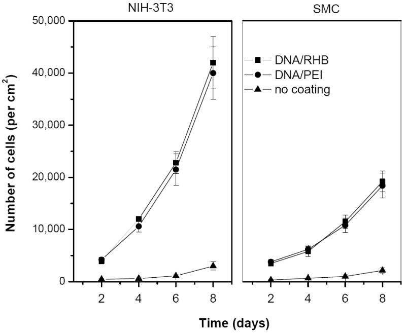
Cell proliferation on stainless steel mesh coated with LbL films. Growth of NIH-3T3 (left panel) and SMC (right panel) was followed by measuring LDH content in cell lysates from the mesh coated with (DNA/RHB)15 (■) or (DNA/PEI)15 (●). Control non-coated mesh (▲). Results are expressed as mean number of cells/cm2 mesh ± SD of triplicate samples.