
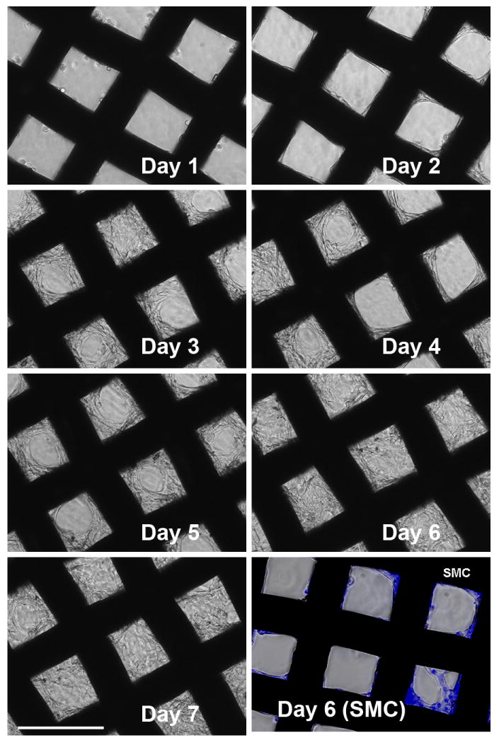
Figure 4.
Cell growth pattern on (DNA/RHB)15 films. NIH-3T3 cells were imaged daily by optical microscopy. Bottom right panel shows SMC imaged 6 days after cell seeding (nuclei stained with Hoechst3342). (size bar = 200 μm)
Official websites use .gov
A
.gov website belongs to an official
government organization in the United States.
Secure .gov websites use HTTPS
A lock (
) or https:// means you've safely
connected to the .gov website. Share sensitive
information only on official, secure websites.
Cell growth pattern on (DNA/RHB)15 films. NIH-3T3 cells were imaged daily by optical microscopy. Bottom right panel shows SMC imaged 6 days after cell seeding (nuclei stained with Hoechst3342). (size bar = 200 μm)