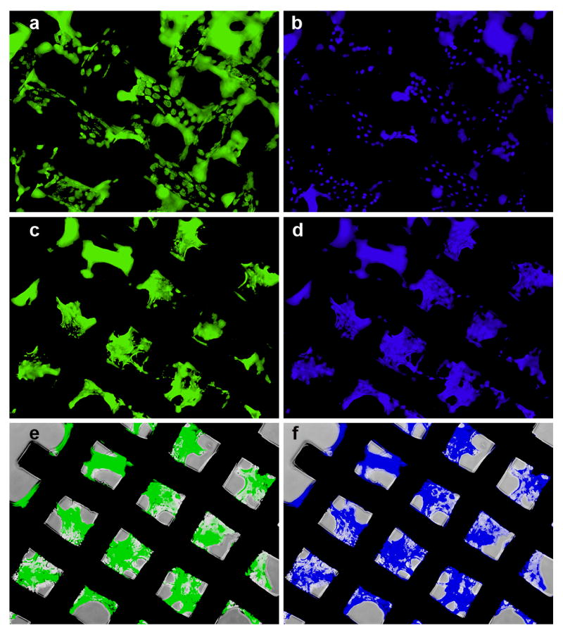
Figure 7.

Transfection activity of (DNA/RHB)15 films based on GFP-DNA. Fluorescence microscopy images of GFP expression in NIH-3T3 cells with the plane of focus on top of the mesh wires (a) and in spaces between the wires (c). Corresponding images of cells with nuclei stained with Hoechst3342 (b, d). Phase and fluorescence overlay (e, f).
