Figure 4.
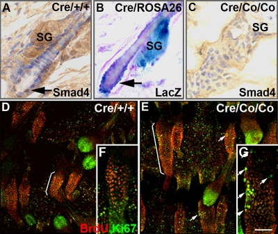
Smad4 deletion result in overactivation of follicle SCs. Immunohistochemistry staining of Smad4 in HFs of wild-type (A) and Smad4 mutants (C). (B) LacZ staining of a HF in a K5-Cre;ROSA26 double transgenic mouse. (D–G) Costaining of BrdU and Ki67 were performed after a 60-d chase. LRCs concentrated in the bulge of wild-type tail epidermis (D and F, brackets), whereas Ki67-labeled cells were concentrated most in the bulb (D and F, arrows). In Smad4 mutants, the LRC zones were expanded (G, brackets), and increased numbers of LRCs were colabeled with Ki67 (E and G, arrows). SG, sebaceous gland. Bar, (A–C) 25 μm; (D and E) 100 μm; (F and G) 40 μm.

