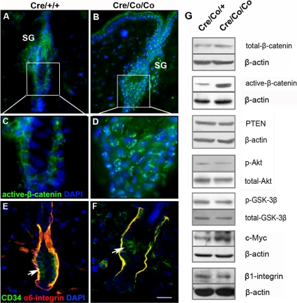
Figure 7.
The increased nuclear localization of β-catenin and increased c-Myc expression in Smad4 mutant epidermis. (A–D) The immunofluorescence of active-β-catenin in the HFs of the control (A and C) and Smad4 mutant mice (B and D) at P42. (E and F) The immunofluorescence of α6-integrin and CD34 in HFs showed that α6-integrin was down-regulated in SC niche of Smad4 mutants at P42. (G) The expression of total β-catenin, active-β-catenin, PTEN, p-Akt, p-GSK-3β, c-Myc, and β1-integrin in epidermis of P42 Smad4 mutant and control mice were detected by Western blot analyses. Bar, (A and B) 35 μm; (C and D) 15 μm; (E and F) 25 μm.

