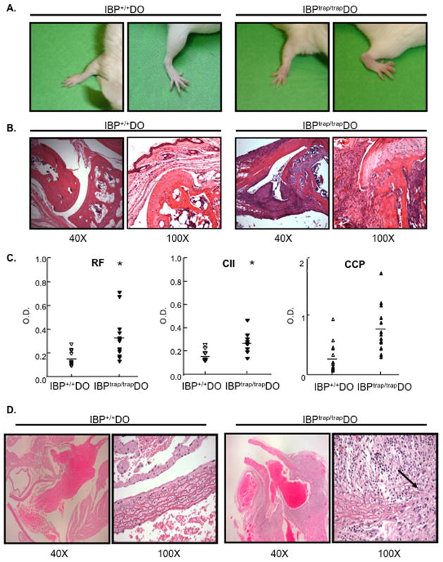
Figure 1.
Development of arthritis and large-vessel vasculitis in IBPtrap/trap DO11.10 mice. A. Shown are the wrist and ankle of a normal 16 wks old IBP+/+ DO11.10 (IBP+/+ DO) female mouse (left) and of an affected IBPtrap/trap DO11.10 (IBPtrap/trap DO) female mouse (right) that had developed swelling and erythema of the joints. B. Histopathologic analysis (hematoxylin/eosin staining) of wrist joints of a 12-week-old female IBP+/+ DO11.10 mouse (left) and an age- and sex- matched IBPtrap/trap DO11.10 mouse (right). Light microscopy images (magnification of 40 and 100×, as indicated) are shown. These findings are representative of 6 mice for each group. C. Serological analysis. Sera from IBP+/+ DO11.10 and IBPtrap/trap DO11.10 mice (6–25 weeks old, male and female, n=6–12) were collected and levels of rheumatoid factor (RF), anti-collagen II (CII) antibodies, and anti-cyclic citrullinated peptide (CCP) antibodies were analyzed by ELISA. *p<0.05. D. Histopathologic analysis (hematoxylin/eosin staining) of the root of the aorta of a 16 wk-old IBP+/+ DO11.10 female mouse (left panels) and an age-matched IBPtrap/trap DO11.10 female mouse (right panels). Light microscopy images (magnification of 40× and 100×, as indicated) are shown. These findings are representative of 6 mice for each group.

