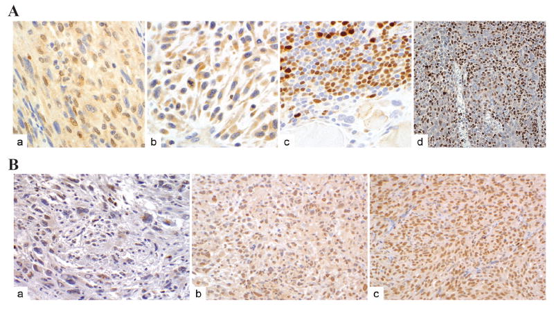
Figure 4. Immunohistochemical analysis of IRF8 and FLIP protein levels in human STS.
Microsections of human STS specimens derived from 60 human STS patients were printed on glass slides as tissue microarray and immune-stained using IRF8-specifc antibody (A) or FLIP-specific antibody (B) as described in Material and Methods. The antibody-specific staining is shown as brown color. A. Four representative specimens with IRF8 staining: in the nuclei of tumor cells (a), cytosolic but no nuclear IRF8 staining of tumor cells (b), lymphoid aggregates in STS specimens showing strong nuclear staining (c), and IRF8 staining in the nuclei of tonsil lymphocytes (d). B. Three representative specimens with scattered low FLIP staining of tumor cells (a), strong FLIP staining of tumor cells (b), and FLIP staining is positive control tissue (c).

