
Figure 6. Ectopic expression of IRF8 sensitized human sarcoma cells to FasL- and TRAIL-induced apoptosis.
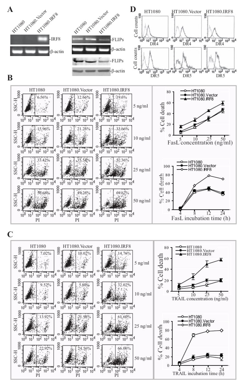
A. RT-PCR analysis of IRF8 expression in non-transfected (HT1080), vector-transfected (HT1080.Vector) and IRF8-transfected (HT1080.IRF8) human sarcoma cells. β-actin was used as normalization standard (left panel). RT-PCR and Western blotting analysis of FLIP expression in HT1080, HT1080.Vector and HT1080.IRF8 human sarcoma cells (right panel). Upper panel is FLIP mRNA level and lower panel shows FLIP protein level. B. Fas-mediated apoptosis in HT1080 sarcoma cell line/sublines. Tumor cells were cultured in the presence of different concentrations of recombinant human FasL for approximately 18 h. The cells were then stained with PI and analyzed by flow cytometry. Shown are histograms of one of three representative experiments (left panel) and plot of FasL concentration against percentage of cell death (top right panel). The tumor cells were also treated with FasL (25ng/ml) and analyzed for cell death at different time points (bottom right panel). C. TRAIL-induced apoptosis in HT1080 sarcoma cell line/sublines. Tumor cells were cultured in the presence of different concentrations of recombinant human TRAIL for approximately 18 h. The cells were then stained with PI and analyzed by flow cytometry. Shown are histograms of one of three representative experiments (left panel) and plot of FasL concentration against percentage of cell death (top right panel). The tumor cells were also treated with TRAIL protein (25ng/ml) and analyzed for cell death at different time points (bottom right panel). D. Cell surface TRAIL receptors DR4 and DR5 expression levels. Tumor cells were stained with anti-DR4- and DR5-specific antibodies, respectively, and analyzed with flow cytometry. Isotype-matched IgG control staining is depicted as gray areas, and DR4- or DR5-specific staining is depicted as solid lines.