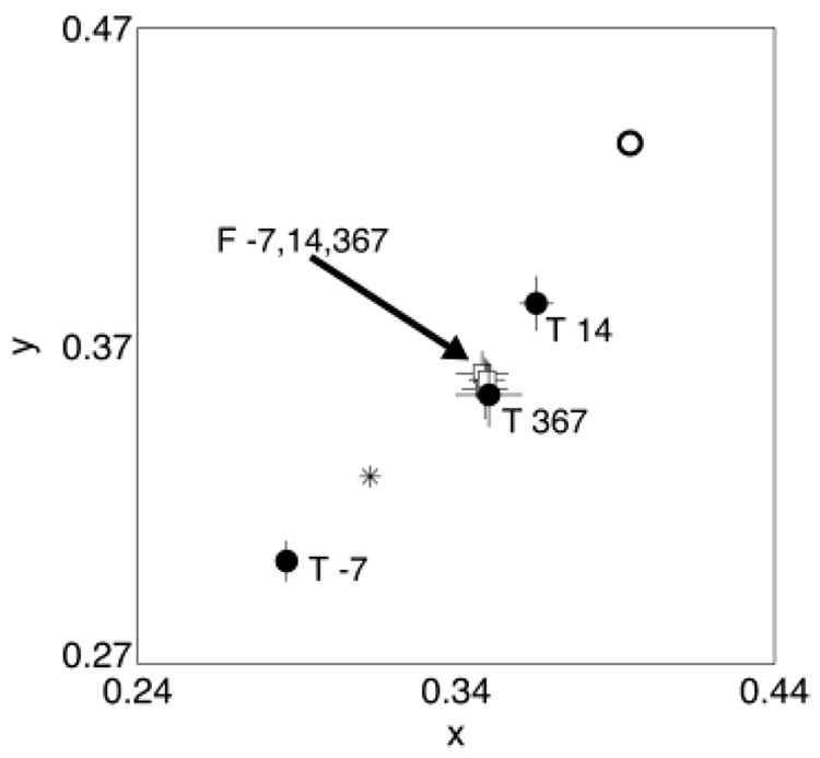
Fig. 5.
Achromatic settings made for observer A using the test (T) eye (closed circles) and the fellow (F) eye (open squares) on three different days relative to surgery. The open circle shows the equivalent presurgery setting calculated using the lens-absorption difference results. The asterisk shows the chromaticity of a typical white point (CIE Illuminant D65). Error bars are ± 1 S.E.M.

