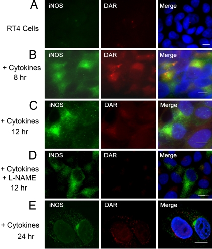
Fig. 4.
Lack of NO in aggresomes of cytokine-induced iNOS. For induction of iNOS, RT4 cells were incubated, for various time points, in the presence or absence of a cytokine mixture of IFN-γ (100 units/ml), IL-1β (0.5 ng/ml), and TNF-α (10 ng/ml). In D, the NOS inhibitor L-NAME was added to culture media. Following cytokine stimulation, cells were incubated for 1 h in the presence of DAR. Cells were then fixed, stained with 4′,6-diamidino-2-phenylindole dihydrochloride (DAPI) to visualize nuclei (blue), and immunolabeled using an iNOS antibody and a goat anti-mouse conjugated to Alexa 488 (green). (Scale bar, 10 μm.)

