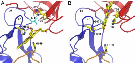
Fig. 3.
Heme-binding after HasA-docking and in a transition state of docking. (A) Heme-binding site in HasA∼HasR∼heme. Both proteins are shown in ribbon representation, HasA in red, the HasR barrel in blue, and the HasR apex C in orange. The axial heme ligands are HasR-His-603 from L7 and HasR-His-189 from apex C of the plug domain. HasR-Ile-671 is shown in cyan. The backbone of the HasA-His-32-bearing loop is seen only partially because of disorder. (B) Heme-binding site in HasA∼HasR-Ile671Gly∼heme. The axial heme ligand is HasA-Tyr-75, hydrogen bonded with HasA-His-83. HasR-Gly-671 is shown in cyan in the backbone of L8. HasA-His-83 in the side-chain conformation seen here would clash with heme in the binding site shown in A, where it is rotated toward HasA. The backbone of the HasA-His-32-bearing loop is seen only partially because of disorder.

