
Figure 4.
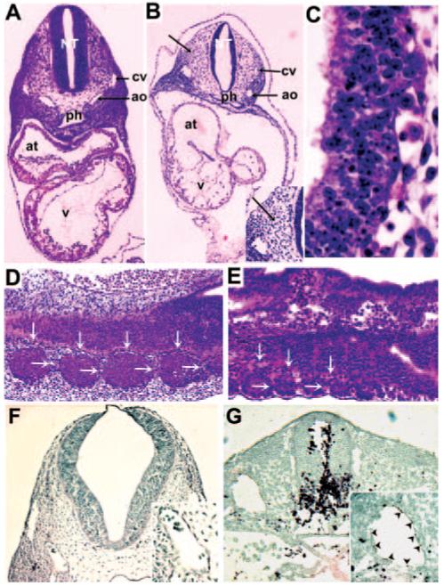
Neuronal and somitic defects in NEKO mice. A and B, Hematoxylin-and-eosin-stained cross sections through day E9.5 embryos of control (A) and NEKO (B) mice; original magnification ×40. Inset shows hypoplastic cardinal vein at higher magnification. C, Higher magnification (×400) of NEKO mice showing pyknotic nuclei in neural tube. D and E, Hematoxylin-and-eosinstained sagittal sections through embryo trunk at day E9.5 of control (D) and NEKO (E) embryos. Arrows denote intersomitic vessels. F and G, Activated caspase-3 staining of control (F) and NEKO (G) mice. Insets represent aortas at higher magnification. ao indicates aorta; at, atrium; cv, cardinal vein; nt, neural tube; ph, pharynx; and v, ventricle.