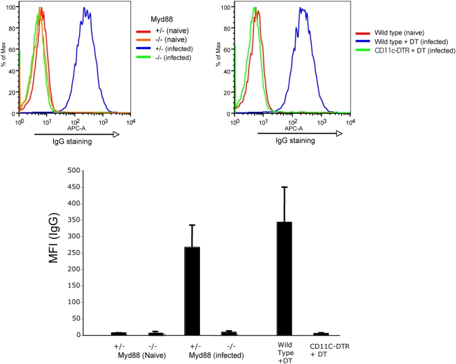
Figure 9. F-MLV–specific IgG is absent from serum of infected mice lacking Myd88 or CD11c+ DCs.
Serum samples from naïve or infected mice were assayed for the presence of F-MLV–specific IgG, by incubation with a suspension of F-MLV–infected cells, followed by secondary staining for mouse IgG. The cells were then analyzed by flow cytometry. Representative stainings for Myd88 knockout mice (upper left) and CD11c+ DC-depleted mice (upper right) are shown. Mean fluorescence intensities (MFI) for the average of five mice were calculated for each condition and plotted (lower panel). The data shown are from one of two independent experiments.

