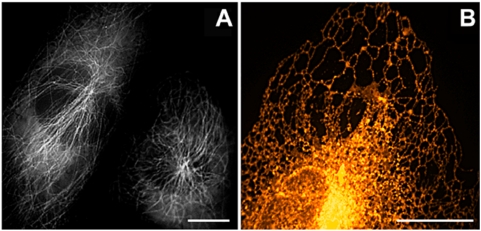
Figure 3. Applications of mRuby as a cellular marker protein.
(A) Epifluorescence image of NIH3T3 cells expressing mRuby fused to human α-tubulin. Two cells from a single image were arranged in closer proximity (B) Spinning-disk confocal image of the endoplasmic reticulum of a HeLa cell stained with ER-mRuby-KDEL. False colors encode fluorescence intensity. Bars: 2.5 µm.

