
Figure 1.
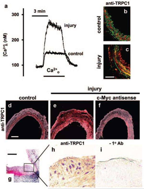
Upregulated TRPC1 in vivo. a, Intracellular calcium concentration ([Ca2+]i) in control and cuff-injured mouse carotid artery. Ca2+o indicates reapplication of extracellular 2.5-mmol/L Ca2+ in the presence of 10 μmol/L verapamil after a 10-minute period in calcium-free solution with 10 μmol/L thapsigargin to deplete intracellular stores. b and c, Immunofluorescence images showing labeling by anti-TRPC1 T1-A antibody (red) in control and parallel cuff-injured mouse carotid artery. The green is autofluorescence from the elastic laminae. The arterial lumen is to the right. The calibration bar in c is 50 μm and applies to b and c. d through f, Immunofluorescence labeling of rat carotid artery with anti-TRPC1 T1-A antibody (red) showing the control contralateral (d) and injured (e) arteries from the same rat and an injured artery from a different rat treated with antisense DNA targeting c-Myc (f). The calibration bar in d is 100 μm and applies to d through f. g, Section of balloon-injured porcine coronary artery stained with hematoxylin and eosin. The calibration bar is 200 μm. h and i, Magnified images of the neointimal plaque shown in g but labeled with (h) or without (i) anti-TRPC1 T1E3 antibody.