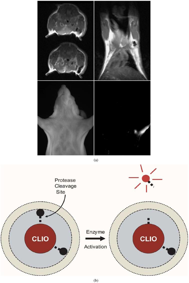
Fig. 1.
(a) Axial and coronal MRI images top and near-infrared fluorescence image bottom right. MRI-optical multimodal particle was injected into the left forepaw 24 h prior to imaging. Draining (sentinel) lymph nodes are readily seen by MRI as well as transcutaneously by fluorescence imaging (from Bioconjug. Chem., vol. 13, no. 3, pp. 554-560, May-Jun. 2002, reprinted with permission). (b) Iron core partially quenches the fluorescence until the linker peptide is enzymatically cleaved by proteases and the fluorochrome is spatially separated from the core. Thus, the fluorescence signal intensity in (a) is proportional to enzyme (protease) activity within the lymph node, providing additional characterization.

