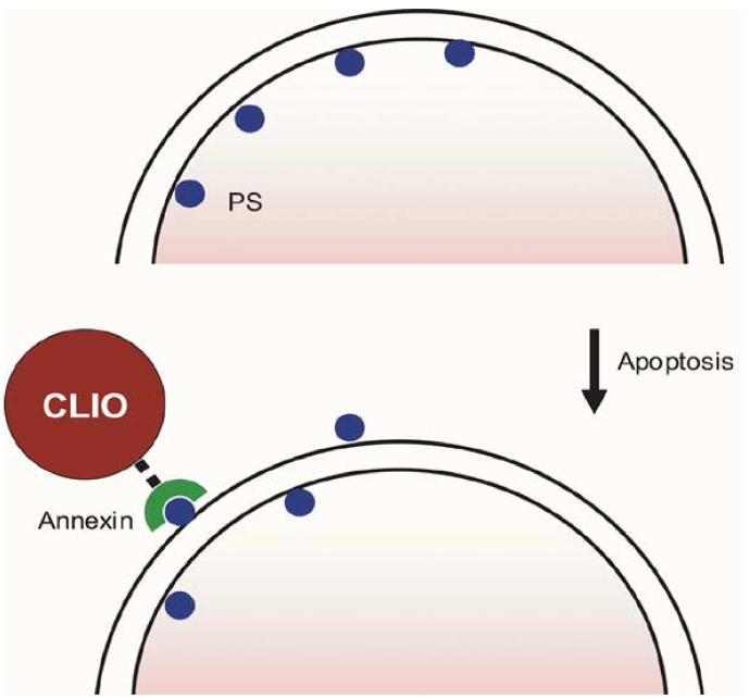
Fig. 3.
Method of MR apoptosis imaging. When cells undergo apoptosis, phosphatidylserine (PS), typically present only on the inner surface of cell membranes, is exposed to the extracellular environment. Annexin V, a human protein with a high affinity for PS, is coupled to an MRI-detectable iron oxide nanoparticle to allow visualization.

