
Figure 1.
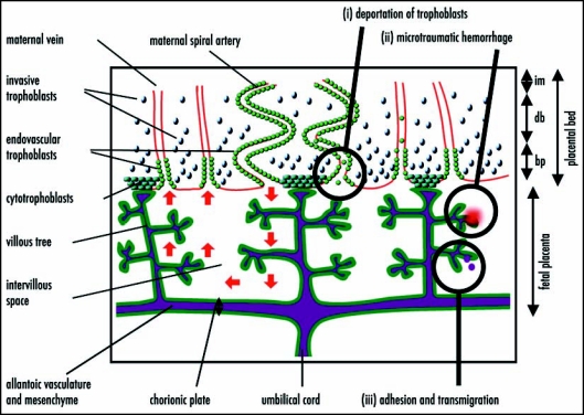
A simplified diagrammatic representation of the structure of the human placenta (adapted from Georgiades et al.36) and hypothesized mechanisms of fetomaternal cell traffic. From the end of the first trimester, maternal blood flows into the fetal placenta via the maternal spiral arteries, through the intervillous space bathing the branches of the villous trees and out through the maternal veins (red arrows on left-hand side). The fetal blood enters via the umbilical cord and circulates to the fetal capillaries in the villous trees. A layer of zygote-derived trophoblasts, in humans a syncytium of syncytiotrophoblasts, on the surface of the villous trees (dark green) forms the barrier between the fetal tissues and the maternal blood. Zygote-derived trophoblasts also progressively invade the placental bed and line the maternal vasculature. By the third trimester the maternal spiral arteries are lined through to the (im), while the maternal veins are lined to the border between the decidua basalis (db) and basal plate (bp). In the mouse, the analogue of the fetal placenta is labyrinthine and the trophoblastic invasion of the maternal blood vessels does not extend beyond the junctional zone analogous to the basal plate. Hypothesized mechanisms of fetomaternal cell traffic include (i) deportation of trophoblasts lining the maternal vessels and intervillous space; (ii) microtraumatic hemorrhage; and (iii) cell adhesion and transmigration across the placental barrier.