
Figure 2.
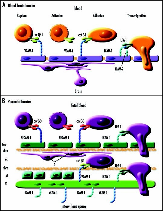
Simplified diagrammatic representations of blood-brain and placental barriers and hypothesized molecular mechanisms of cell adhesion and transmigration. (A) A simplified diagrammatic representation of multistep lymphocyte recognition and capture from blood at the blood brain barrier (adapted from Engelhardt48). Cells expressing α4β1 are captured by VCAM-1 expressed by endothelial cells. There is a rapid activation phase (seconds) that may involve lymphoid chemokines CCL19/ELC and CCL21/SLC. There is a prolonged adhesion phase (hours) followed by slow transmigration (hours) dependent upon binding of LFA-1 to ICAM-1 and/or ICAM-2 on the endothelial cells. It is hypothesized that a similar molecular mechanism may explain fetal cell migration across the blood-brain barrier and the placental barrier. (B) A simplified diagrammatic representation of the human placental barrier showing a hypothetical mechanism of fetal cell capture, adhesion and transmigration. The placental barrier comprises of fetal capillary endothelial cells (fcec), an endothelial basement membrane (ebm), the villous core (vc) which at some interfaces contains pericytes (p) and extracellular matrix, a trophoblastic basement membrane (tbm), in the first trimester a layer of proliferative cytotrophoblasts (ct), and a multinucleated syncytium of syncytiotrophoblasts (ss). In the mouse, the trophoblastic layers differ in that there are two syncytiotrophoblastic layers and the cytotrophoblastic layer is outermost facing the intervillous interface. It is hypothesized that fetal cells may adhere and transmigrate across the placental barrier in a similar manner to that by which lymphocytes cross the blood-brain barrier.