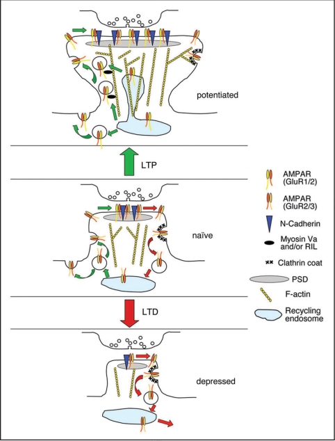
Figure 2.
Relationship between AMPA receptor trafficking, actin dynamics and spine size during synaptic plasticity. Under basal conditions, AMPARs undergo successive rounds of endocytosis, recycling and reinsertion. Spine structure is supported by the underlying actin cytoskeleton. This may in part be maintained by the GluR2/N-Cadherin complex at the PSD. A balance between endocytosis and exocytosis preserves the spine plasma membrane area. During LTP, actin polymerisation is enhanced, resulting in increased F-actin in spines, enlarging the spine head. These actin filaments may provide transport routes for GluR1-containing AMPARs trafficking towards the plasma membrane, via myosin Va or RIL. A shift towards actin polymerization also reduces AMPAR endocytosis. The increase in surface AMPARs leads to spine enlargement via the GluR2/Cadherin complex, possibly by signaling to the actin cytoskeleton. The increase in AMPAR insertion from recycling endosomes contributes additional membrane to the surface of the growing spine. During LTD, a shift towards actin depolymerization results in less F-actin and hence a smaller spine head. Therefore, fewer actin filaments are present to support actin-based trafficking towards the plasma membrane. Actin depolymerization promotes PICK1-mediated AMPAR internalisation. The reduced surface GluR2 contributes to spine shrinkage, possibly via reduced signaling from N-Cadherin. The increase in AMPAR internalization may also result in a net internalization of plasma membrane, reducing the surface membrane of the shrinking spine.

