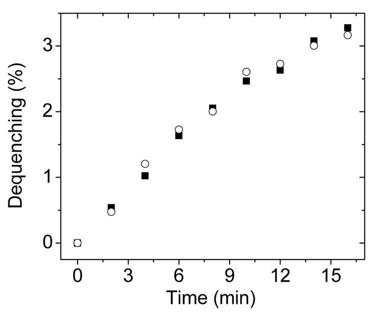
Fig. 2.
Results from two different R18 dequenching assays. In the first experiments (filled squares) only donor vesicles and GM2AP were mixed. Spectra were recorded for GM2AP final concentration of 0.75 µM with 20 µM POPC:R18 (95:5) vesicles. In the second experiments, (open circles) donor vesicles, GM2AP and acceptor vesicles were mixed. Here, 20 µM POPC:R18 (95:5) donor vesicles and 20 µM POPC vesicles were incubated for 10 min; GM2AP was added to a final concentration of 0.75 µM. Before addition of GM2AP, the emission intensity at 590 nm was set as 0% dequenching. The intensities after complete solubilization of the POPC:R18 (95:5) vesicles by 0.1% Triton-100 was set as 100% dequenching. Spectra collected with 570 nm excitation wavelength.

