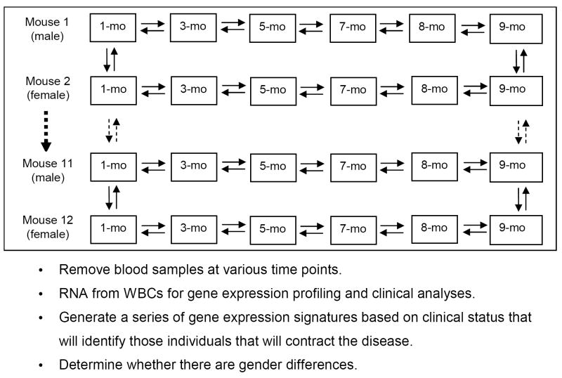
Fig. 1.
Microarray experimental design to predict disease. Each box represents a time point for a given AKR/J mouse. The arrows between the boxes denote duplicate microarrays with a dye flip. The larger dashed arrow represents mice 3 through 10, and the smaller dashed arrows represent the microarray comparisons carried out at months (mo) 1 and 9 (the beginning and end of the study) between mice 1 and 2, mice 2 and 3, etc., to mice 11 and 12. The odd numbered mice were male and the even-numbered mice were female.

