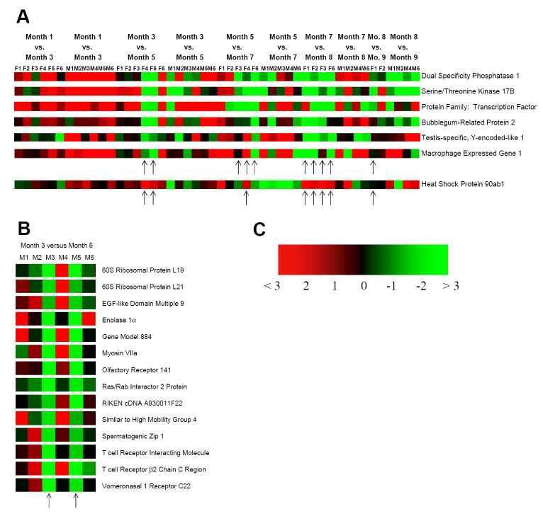
Fig. 5.
Gender-specific predictor gene sets for lymphatic leukemia in the AKR/J mouse. WBCs were collected from mice at 1, 3, 5, 7, 8, and 9 months of age. Heat maps (p-value ≤ 0.05) of those genes predicting the onset of lymphatic leukemia are shown for the female (F) AKR/J mice (A) and the male (M) AKR/J mice (B). The upward-pointing arrows denote the predictor genes displayed at the different ages for each mouse. The color gradient in (C) is an indicator of the relative mRNA levels in the heat map, in which green and red indicate lower and higher fold differences, respectively.

