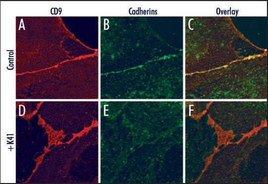
Figure 1.
Cell surface localization of cadherins and CD9. Vero cells were not (A–C), or were (D–F) preincubated with 10 µg/ml mAb K41 for 2 h at 37°C. Expression of CD9 was detected using mAb K41 and secondary Alexa-594-conjugated antibodies (red, A and D). Expression of cadherins was detected using pan-cadherin antibodies (Cell Signaling Technology) and secondary Alexa-488 antibodies (green, B and E). The overlay (C and F) reveals a colocalization of cadherins and CD9 predominantly in the absence of K41-preincubation at the cell border (C). The larger CD9-positive net-like structures at cell contact areas as detected in (D) do not contain enriched amounts of cadherins.

