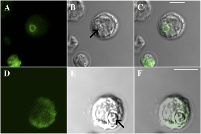
Figure 1.
Immunofluorescent localization of LCIB in C. reinhardtii L-CO2-acclimated CC125 cells. A and D, False color immunofluorescence images of two different cells. B and E, Confocal images of the same cells. C and F, Merged images. The white bars in C and F are 5 μm in length and the arrows in B and E indicate the location of the pyrenoid in each confocal image.

