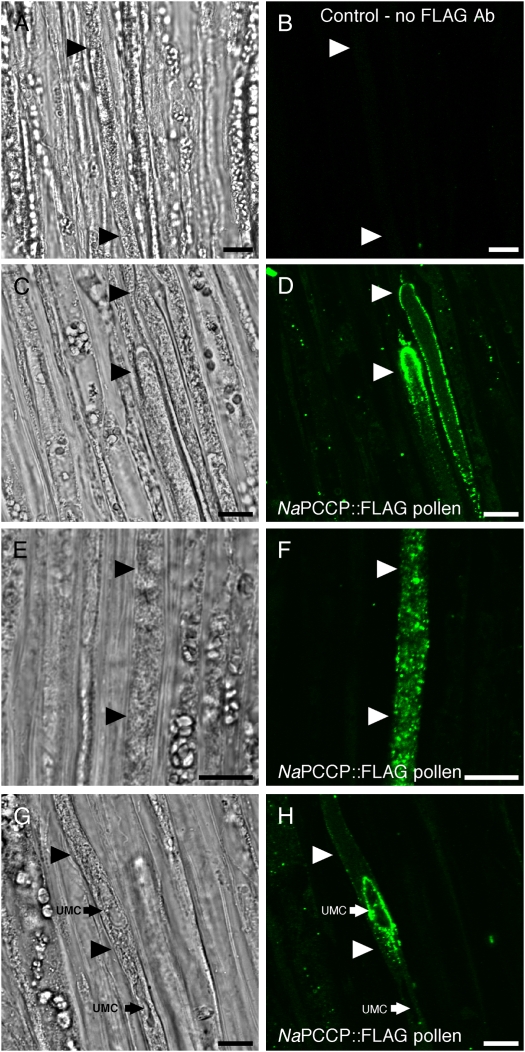
Figure 4.
NaPCCP∷FLAG is localized to pollen tube membranes. N. tabacum pistils were pollinated with NaPCCP∷FLAG pollen and prepared for immunohistochemistry after 18 h. Bright-field (left) and fluorescence (right) images are shown; arrowheads show pollen tubes. A and B, Control; no FLAG antibody. C to H, Representative NaPCCP∷FLAG localization patterns. C and D, Tip localization seen in approximately 80% of pollen tube tips. E and F, Punctate pattern seen behind the tip in approximately 50% of pollen tubes. G and H, Localization around a UMC (small arrow) seen in approximately 30% of pollen tubes. Bars = 10 μm. [See online article for color version of this figure.]

