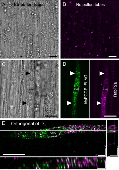
Figure 7.
Largely independent localization of NaPCCP∷FLAG- and anti-RabF2a-labeled endosomes. Unpollinated control (A and B) or N. tabacum pistils pollinated with NaPCCP∷FLAG pollen (C–E) were immunostained with anti-FLAG or anti-RabF2a (endosomal marker) antibodies. The unpollinated control images (A and B) show RabF2a-positive endosomes in transmitting tract cells. C, Bright-field image showing a NaPCCP∷FLAG pollen tube approximately 500 μm behind the tip. D, Single optical section of C showing a pollen tube with a prominent vacuole. Left, Anti-FLAG (green); right, anti-RabF2a (magenta). E, Orthogonal views of a deconvolved, merged stack from C and D. The main panel shows a single optical section (x-y plane); cross-hairs indicate where the stack was sectioned to show the y-z and x-z planes. White spots indicate rare areas of colocalization, but the NaPCCP∷FLAG (green) and RabF2a (magenta) signals are largely independent. Bars = 10 μm.

