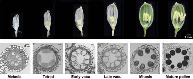
Figure 1.
Photographs of spikelets and cross sections of anthers of six progressive developmental stages. Top row (dissecting microscopy), Spikelets with one side of the spikelet brackets removed to reveal the internal anthers. Bottom row (light microscopy), Cross sections of one lobe of an anther of each developmental stage, named according to the morphological features in the microspores. The arrow locates the tapetum layer enclosing the microspores in the locule. In the mature-pollen stage, the tapetum layer had disintegrated. In the bottom row, scale bar = 10 μm; its diminishing length in the images of the six progressive stages reflects the continuous enlargement of the anthers and microspores. vacu, Vacuolated.

