
Fig. 6.
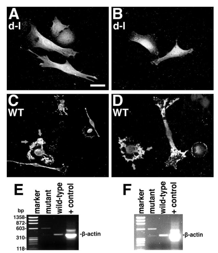
A-D, in situ hybridization of homozygous null mutant (A, B) and wild-type control (C, D) PNS cells with a fluorescent probe for β-actin mRNA; arrows denote the high concentration of β-actin mRNA at the leading edge. Bar = 20μm. (E, F) RT-PCR of anti-myosin-Va immunoprecipitates from homozygous null mutant and wild-type control primary adipose fibroblasts. In F, the levels have been increased to show the faint, but expected band in the mutant control lane, as well as the absence of background bands in the wild-type experimental lane, demonstrating the absence of contamination during gel loading from the positive control lane (RT-PCR from total HeLa cell RNA). The marker lane is PhiX174 RF DNA cleaved with HaeIII.