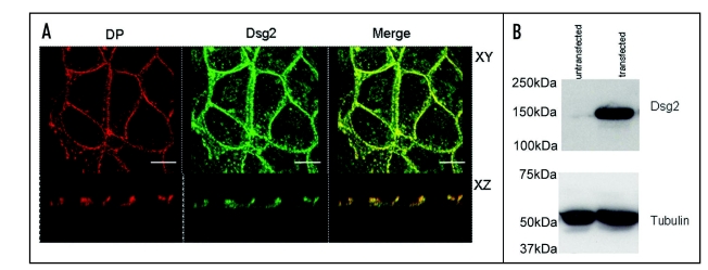
Figure 2.
Exogenous Dsg2 colocalizes with DP and is strongly expressed. (A) Confocal images of MDCK cells with stable expression of mDsg2HA stained for DP with monoclonal antibody 11–5F (left) and polyclonal anti-HA antibody (centre). The merged image (right) shows that Dsg2 is colocalized with DP in both the XY and XZ planes. (B) Immunoblots of equally loaded gel (tubulin control) showing strong expression of mDsg2HA detected with polyclonal anti-HA antibody. Bars = 5 µm.

