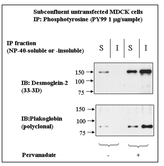
Figure 5.
Dsg2 and Pg are slightly tyrosine-phosphorylated in the insoluble fraction of control cells. Untransfected MDCK cells were either untreated or treated with pervanadate for 15 minutes prior to detergent fractionation and subsequent immunoprecipitation using the monoclonal anti-phosphotyrosine antibody PY99. Samples were fractionated with equal loading by SDS-PAGE and immunoblotted for endogenous Dsg2 and Pg. As expected Dsg2 and Pg in the pervanadate-treated samples were tyrosine-phosphorylated. There was also tyrosine-phosphorylated Dsg2 and Pg in the soluble fraction of untreated cells yet apparently none in the insoluble fraction. As this population of protein was undetectable within the ectopic mDsg2HA IPs it must be assumed that it represents a small proportion of the cellular pool of these proteins.

