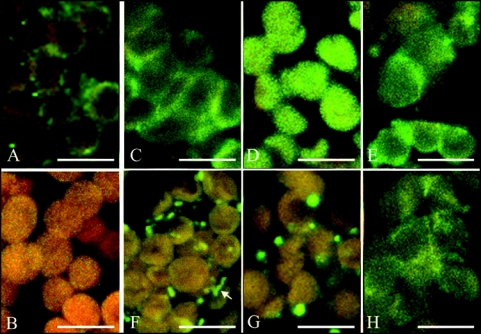
Figure 1.
Confocal immunofluorescence images of Arabidopsis thaliana mesophyll, wild type (WT), phot1 and phot2 mutant plants. Myosins are labeled with anti-rabbit (smooth and skeletal) antibodies and with secondary FITC-conjugated antibodies (yellow-green color). Red-orange color comes from chloroplast autofluorescence. Left column, WT, Arrangements of myosins in the dark-adapted tissue (A); Control with secondary antibodies only (B); Wild type cells, weak blue light-irradiated (C). Wild type tissue, strong blue light-irradiated (F). Weak (D) and strong (G) blue light-irradiated phot1 mutant. Weak (E) and strong (H) blue light-irradiated phot2 mutant. Note the difference in the arrangements of myosins between strong and weak blue-irradiated WT tissue and the similarity of phot2 irrespective of light intensity. The arrow at (F) points at myosins located along an actin cable. The bars are 10 µm.

