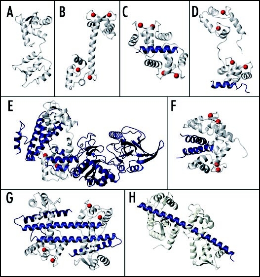
Figure 1.
Structures of CaM and CaM-target complexes. (A) apo-CaM (PDB:1DMo), (B) Ca2+-CaM (PDB:1CLL). Complexes of CaM bound to (C) CaMBD of smooth muscle myosin light chain kinase (PDB:1CDL), (D) partial CaMBD of plasma membrane Ca2+-pump C20W (PDB:1CFF), (E) the adenylyl cyclase protein from Bacillus anthracis (PDB:1K93), (F) 2 glutamate decarboxylase CaMBD's (PDB:1NWD), (G) 2 CaM proteins bound to 2 small conductance Ca2+-activated potassium channel (SK channel) CaMBD's (PDB:1G4Y), (H) 2 apo-CaM proteins bound to 2 tandem IQ motifs from murine myosin V (PDB:2IX7). In each panel CaM is shown in ivory, the target molecule is shown in blue and the Ca2+ ions bound to the N- and/or C-lobes of CaM are represented by red spheres.

