
Figure 3.
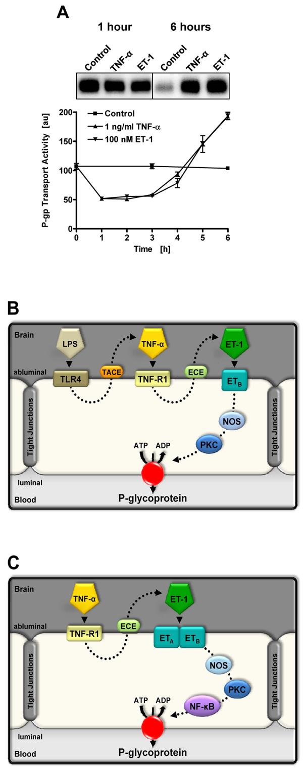
Proinflammatory signaling to P-glycoprotein in rat brain capillaries. (A) Time course showing changes in P-glycoprotein-mediated transport activity in isolated rat brain capillaries exposed to TNF-α and ET-1. Note the rapid reduction in activity and the delayed increase over control levels. The inset shows Western blots of capillary membranes after 1 h and 6 h of exposure. Note the absence of change in P-glycoprotein expression after 1 h and the dramatic change after 6 h (Bauer et al., 2007a). These blots were processed using different exposure times and expression levels cannot be compared across blots. We showed constant P-glycoprotein expression in capillaries over 6 h of incubation in control medium (Bauer et al., 2007a). (B) LPS, TNF-α and ET-1 signaling to P-glycoprotein in the short-term (rapid reduction of transport activity (Hartz et al., 2004; 2006)). (C) TNF-α and ET-1 signaling to P-glycoprotein in the long-term (increased transport activity and transporter expression (Bauer et al., 2007a)).