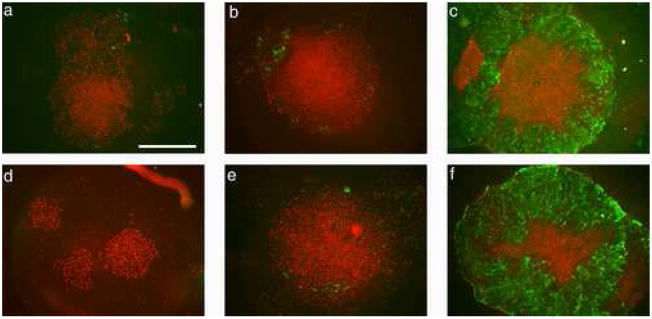
FIGURE 5.
H1 hESC cultured in the presence of BMP4 for 1 (a, d), 3 (b, e), or 5 days (c, f) in either low (L) (a–c) or atmospheric (A) (d–f) oxygen immunostained for Oct4 and SSEA-1. By d 5, immunostaining for Oct4 (red) becomes confined to an inner core of densely packed cells, but is still associated with nuclei of most cells in the colonies at d 1 and 3. Note that in d, which shows three small colonies, all the cells have apparently responded to BMP4 but all the nuclei remain Oct4-positive. By contrast, staining for the differentiation marker SSEA-1 (green) is initially confined to a few outer cells at d 3 (b, e), but becomes associated with all cells outside the dense central core of Oct4-positive cells by d 5 (c, f) under both oxygen conditions. Although, not shown, some smaller colonies were entirely SSEA1-positive and Oct4-negative by d 5. Whole colonies were imaged on the Olympus epifluorescence system under a 4x objective. The scale bar in a represents 1mm.

