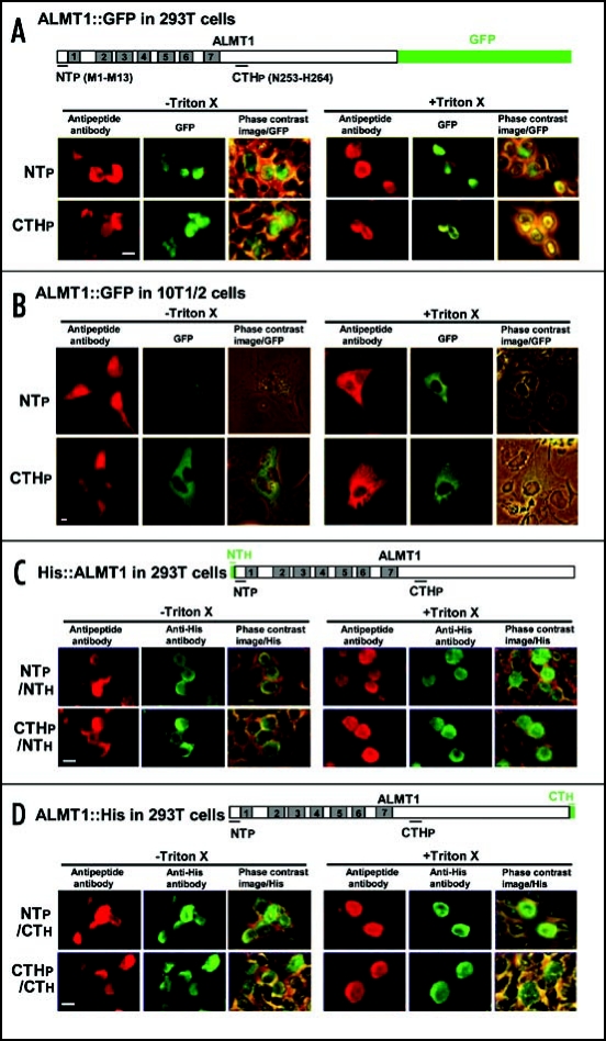
Figure 2.
The orientation of the N- and C-termini of ALMT1 expressed in mammalian cells (293T, 10T1/2). Cells transiently expressing the ALMT1 construct fused with GFP at the C-terminus (A and B), tagged with a His epitope at the N-terminus [NTh, (C)] or tagged with a His epitope at the C-terminus [CTh, (D)] were identified with green GFP fluorescence and the green AF488 fluorescence obtained with anti-His antibody, respectively, in the presence or absence of Triton X-100. These cells were simultaneously tested for the accessibility of the polypeptide epitopes located at the N-terminus (NTp) and C-terminal half region (CTHp) to their respective anti-peptide antibodies (detected by red AF594 fluorescence). Representative images are shown here. Left panels, AF594 fluorescence; Center panels, GFP fluorescence or AF488 fluorescence; Right panels, the phase contrast image overlaid with the GFP or AF488 fluorescence. Bar = 20 µm.

