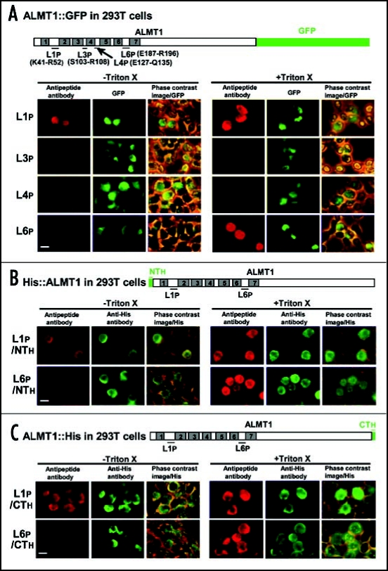
Figure 3.
The orientation of putative loop regions (L1, L3, L4, L6) of ALMT1 expressed in 293T cells. Cells transiently expressing the ALMT1 construct fused with GFP at the C-terminus (A) (detected by GFP fluorescence) or tagged with His at the N-terminus (B) or C-terminus (C) (detected by AF488 fluorescence) were identified as described in Figure 2, in the presence or absence of Triton X-100. These cells were simultaneously tested for the accessibility of the polypeptide epitopes located at L1 (L1p, shown with the amino acid positions of the ends of the epitope) and L6 (L6p) to their respective anti-peptide antibodies (detected by red AF594 fluorescence). Left panels, AF594 fluorescence; Center panels, GFP fluorescence or AF488 fluorescence; Right panels, the phase contrast image overlaid with the GFP or AF488 fluorescence. Bar = 20 µm.

