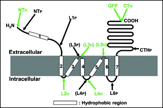
Figure 5.
Topological model of ALMT1. The model depicts the membrane topology of ALMT1 as determined by experimental evidence derived from the accessibility of antibodies to the epitopes (shown with arrows) when ALMT1 is expressed in mammalian cells (Figs. 2–4). The ALMT1 protein consists of hydrophobic N-terminal half and hydrophilic C-terminal half regions. The C-terminal half region (240 amino acids including CTHp) is extracellular. The N-terminal region, including the N-terminus, putative transmembrane domain 1 (TMD 1) and putative loop 1 (L1), is extracellular. Six TMDs follow (2 to 7) and three loops (L2, L4, L6) faced the cytsol. L4 was not detected by antipeptide antibody (shown in parentheses) but was detected with an anti-His antibody. However, both L3 and L5 were not detected even with the anti-His antibody (L3 was not detected by antipeptide antibody either and is shown in parentheses), suggesting that these loops are too small to be detected by the antibodies or could be embedded inside the membrane.

