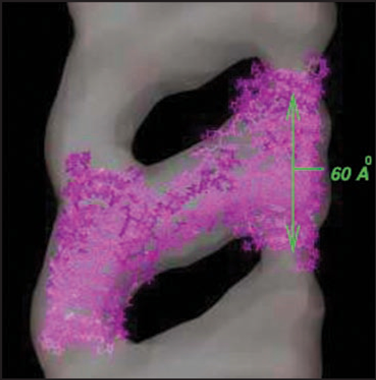
Figure 1.

30 Å resolution electron density map of in vitro grown mammalian prion protein fibril from ref. 9 (grey) with model repeat unit of four PrP proteins adopting beta helical conformations in both the N-terminal and C-terminal regions embedded (lavender). Embedding of the tetramer model and image production through Chimera.65
